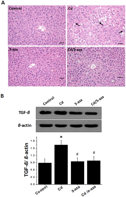
Figure 6. Effects of chronic exposure to Cd on the pathological changes and TGF-β expression in mouse livers.
Liver tissues collected as described in Figure 4 were used for pathological examination using H&E staining (A), which showed significant increases in lipid drops (vacuoles) with infiltration of inflammatory cells (arrows), and for Western blotting of TGF-β expression (B). Bar = 50 µm. Data was presented as mean ± SD (n = 10). * p<0.05 vs control, # p<0.05 vs Cd group.

