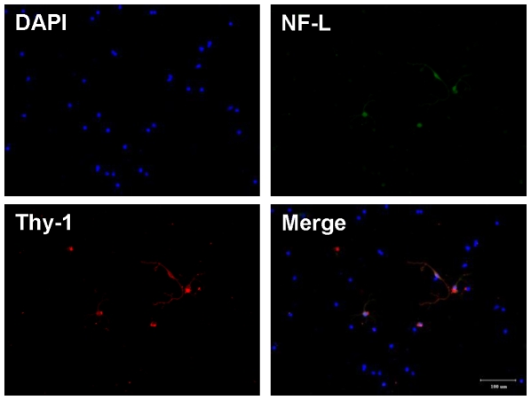
Figure 4. Fluorescence images of dissociated RGCs.
Retinal cells were dissociated with papain, fixed with 4% paraformaldehyde in PBS, and stained with DAPI (blue). RGCs (arrowhead) were identified by double immunocytochemistry with anti-neurofilament (NF)-L antibody (green) and anti-thy-1 anitbody (red), bar = 100 µm.

