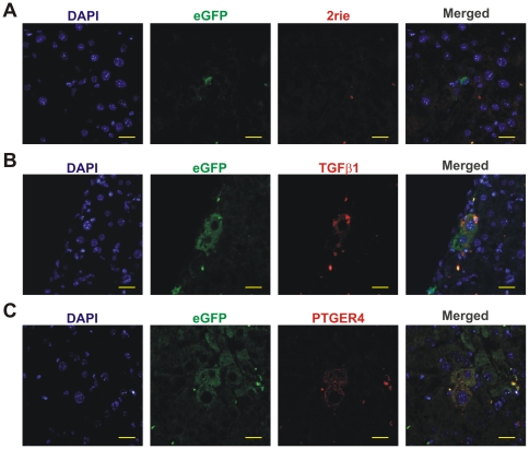
Figure 4. Tgfβ1 and Ptger4 are overexpressed in BMDH.
A. Hepatic section sequentially stained, as described in Materials and Methods, for eGFP detection and with the secondary antibody donkey anti-rabbit-TexasRed. B. Tgfβ1 expression in BMDH. C. Ptger4 presence in BMDH. 20 µm scale bars are shown.

