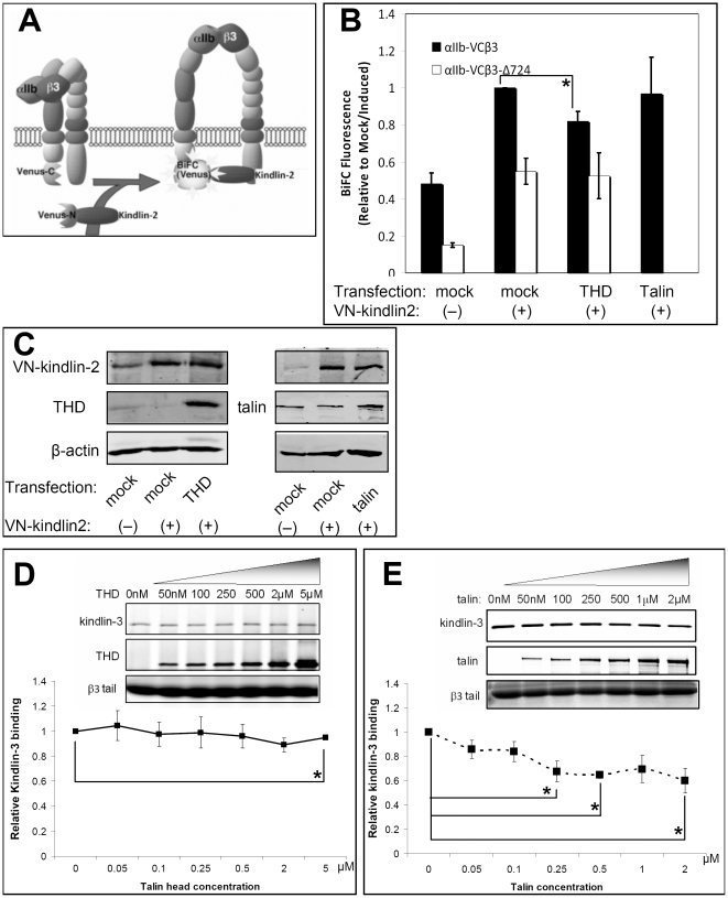
Figure 6. Effect of talin or THD on interaction between kindlins and αIIbβ3.
(A) Schematic illustration of BiFC in CHO cells depicting αIIb-VC, VN-kindlin-2 and β3. When VN-kindlin interacts with αIIb-VCβ3 through the β3 tail, VN and VC should reconstitute Venus, resulting in BiFC. (B) Over-expression of THD or talin does not promote kindlin-2 interaction with αIIbβ3. CHO cells expressing αIIb-VCβ3 or αIIb-VCβ3Δ724 was co-transfected with Tac as a transfection marker and an expression vector for THD, talin or empty vector (Mock), as indicated. After induction of VN-kindlin-2 expression with doxycycline, BiFC was quantified by flow cytometry. BiFC fluorescence was normalized to αIIbβ3 expression and expressed as fold-increase relative to doxycycline-treated, Mock-transfected cells. Data represent means ± SEM of three experiments (asterisk denotes statistically significant difference against mock/induced, P<0.05). (C) Western blots were performed to monitor expression of VN-kindlin-2, talin and THD in cell lysates. (D) Purified kindlin-3 with or without addition of THD was incubated with the recombinant β3 cytoplasmic tail conjugated to neutravidin beads. After washing, proteins bound to the beads were detected on western blots. Band intensities were quantified in LICOR, normalized to kindlin-3 binding in the absence of THD, and presented as a curve. Insert shows a representative western blot of 3 independent experiments. Increasing amount of β3 tail bound THD failed to promote β3–kindlin-3 interaction. Data represent means ± SEM of three experiments. (E) Similar to (D) but talin was used instead of THD. Increasing amounts of β3 tail-bound talin failed to promote β3–kindlin-3 interaction. (asterisks in D and E denotes statistically significant differences compared to kindlin binding in the absence of talin or THD, P<0.10).

