
Figure 2.
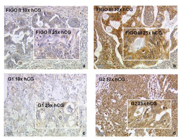
Representative slides of immunohistochemical staining for hCG expression for FIGO stage II (a, hCG negative), FIGO stage III (b, hCG positive) for grade 1 (c, weak hCG staining) and grade 2 (d, strong hCG staining) ovarian cancer tissue. No hCG immunoreactivity was detected in tumor stroma (magnification 10× and 25×).