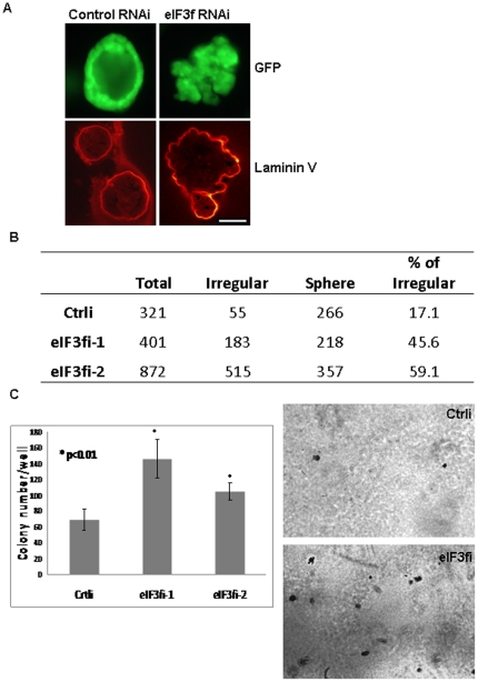
Figure 3. eIF3f-silenced HPDE cells showed malignant features in 3D culture and soft agar assay.
(A) Control and eIF3f-silenced HPDE cells were stably transduced with a GFP-expressing lentivirus (pLKO-puro CMV-TurboGFP, Sigma-Aldrich) and positive cells were sorted by a cell sorter (FACSAria) (Fig. S3). These GFP-expressing cells were used in an ex vivo 3D- cell culture system, immunofluorescent and confocal microscopy analysis. Laminin V (indicating basement membrane) was labeled with laminin V antibody and secondary antibody conjugated with Texas Red. A representative picture of our 3D cell culture system is shown. Note the loss of normal architecture, of cellular polarity, and of smooth basement membrane in eIF3f-silenced HPDE cells. Bar: 50 µm. (B) We counted regular sphere and irregular structures of control cell lines and 2 eIF3f-silenced HPDE cell lines after 10 days of 3D culture. Note that eIF3f-silenced cells formed much more irregular structures than control cells. (C) Soft agar assay: control and eIF3f-silenced HPDE cells were seeded at a density of 10000 cells per well in 6-well plate in 2 ml 0.33% agar and cultured for 14 days. Colonies were stained with 0.05% crystal violet overnight at 4°C. Colonies in the entire well were counted. Representative images of colonies and histogram is shown.

