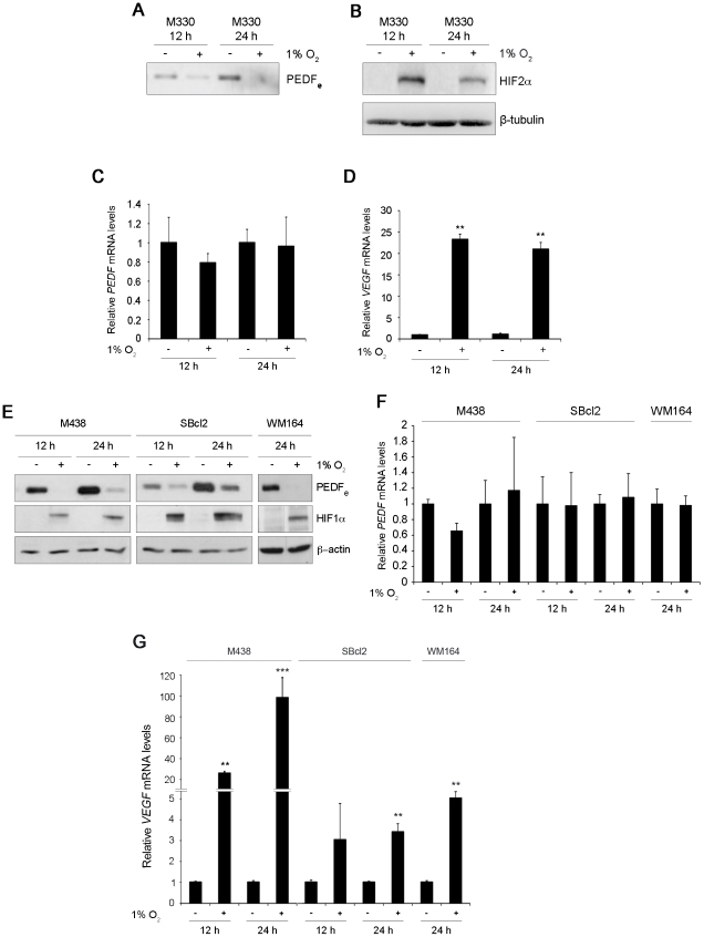
Figure 1. Hypoxia downregulates PEDF at the protein level in melanocytes and human melanoma cell lines.
Western blot analysis of (A) extracellular PEDF (PEDFe) protein levels in conditioned medium (CM) and (B) HIF2α protein levels in whole-cell extracts (B) from M330 primary melanocytes incubated under normoxia (21% O2) or hypoxia (1% O2) for 12 h or 24 h. β-tubulin was used as loading control. Quantitative RT-PCR analysis of (C) PEDF mRNA levels and (D) VEGF mRNA levels in M330 primary melanocytes incubated in normoxia or hypoxia for 12 h or 24 h. PEDF and VEGF mRNA levels are shown relative to cells in normoxia after normalization to β-actin. Bars represent average ± standard deviation (SD) (**P<0.01). (E) Western blot analysis of PEDFe protein levels in CM and HIF1α in whole-cell extracts from M438 primary melanocytes, SBcl2 and WM164 melanoma cell lines incubated in normoxia or hypoxia for 12 h or 24 h. β-actin was used as loading control. Quantitative RT-PCR analysis of (F) PEDF mRNA levels and (G) VEGF mRNA levels in M438 primary melanocytes, SBcl2 and WM164 melanoma cell lines incubated under normoxia or hypoxia for 12 h or 24 h. PEDF and VEGF mRNA levels are shown relative to normoxia after normalization to β-actin. Bars represent average ± SD (**P<0.01; ***P<0.001).

