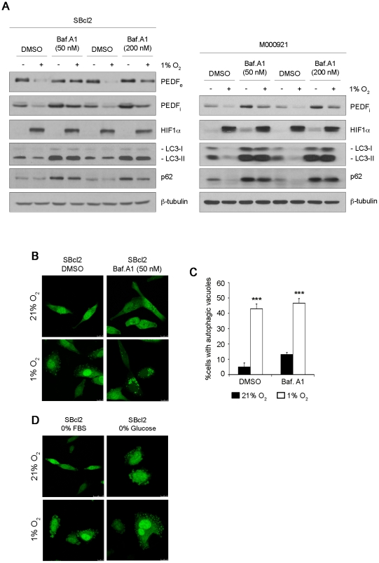
Figure 6. Autophagy is involved in downregulation of PEDF by hypoxia in melanoma cells.
(A) Western blot analysis of extracellular PEDF (PEDFe) protein levels in 24 h conditioned medium (CM), intracellular PEDF (PEDFi), HIF1α LC3 and p62 protein levels in whole-cell extracts from SBcl2 (left) and M000921 (right) melanoma cell lines treated with different concentrations of the autophagy inhibitor bafilomycin A1 (Baf. A1, 50 nM and 200 nM) or DMSO vehicle under normoxic (21% O2) or hypoxic (1% O2) conditions. β-tubulin was used as loading control. (B) Fluorescence images (63× magnification) of GFP-LC3 protein redistribution in SBcl2 melanoma cell line (transduced with pLV-EGFP-LC3 plasmid) treated with 50 nM Baf. A1 in normoxia or hypoxia for 24 h. (C) Quantification of SBcl2 cells with autophagic vacuoles after Baf. A1 treatment for 24 h under normoxia (filled bars) or hypoxia (empty bars). Ten fields from each condition were counted for quantification. Bars represent average ± standard deviation (SD) (***P<0.001). (D) Fluorescence images (63× magnification) of GFP-LC3 redistribution in SBcl2 melanoma cell line grown in the absence of growth factors (0% FBS) or ischemic conditions (0% glucose) under normoxia or hypoxia.

