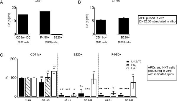
Fig. 7. αGC acC8 can be presented by DCs to induce IL-4, IFN-γ and IL-12p70.
(A) CD11chi CD8α+ DCs, B220+ B cells, and CD11clo CD11blo F4/80+ macrophages were sorted from pooled spleens from 3 B6 mice 2 h after injection of 2 μg αGC and cocultured at indicated numbers with the hybridoma DN32.D3 (50,000 cells/well) overnight and IL-2 released in the supernatant was measured. Error bars represent SEM (B) CD11c+ DCs and B220+ B cells were enriched using autoMACS from the pooled spleens of 3 B6 mice 2 h after injection of 50 μg αGC acC8 and cocultured at indicated numbers with DN32.D3 as in (A). (C) CD11chi DCs, B220+ B cells, CD11clo CD11blo F4/80+ macrophages were sorted from the spleens of uninjected mice and cocultured at 5000 cells per well in the presence of 1 μg/ml αGC or 2.5 μg/ml αGC acC8 for 48 hours with sorted CD5+ cells (50,000 per well) obtained from the spleens of Vα14 Tg mice. IL12-p70, IFN-γ and IL-4 were measured in supernatants. Three separate experiments were performed and data were normalized to the DC+αGC group (100%) in each experiment. Error bars indicate SEM.

