Figure 6.
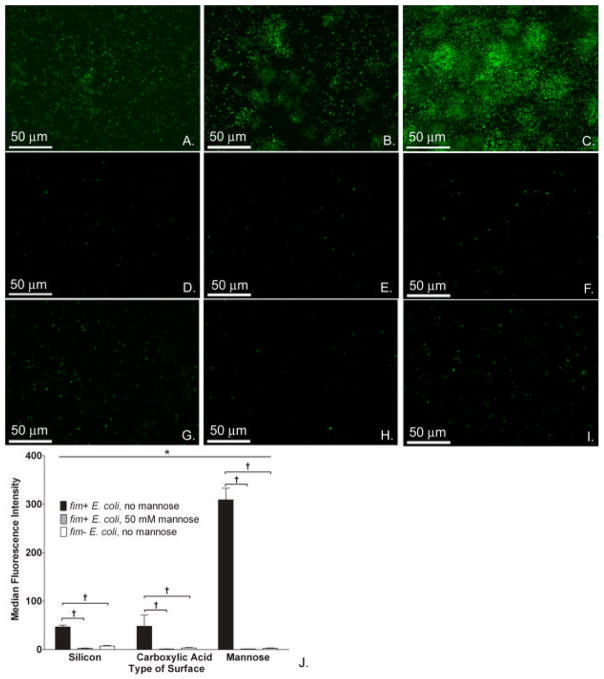
Type 1 fimbriae-mannose interaction is key to enhancement of E. coli biofilm formation on mannose surfaces. Fluorescence images of fim+ E. coli 83972 (BWT8, green) on silicon (A), carboxylic acid (B), and mannose (C) surfaces in the absence of mannose in media. Fluorescence images of fim− E. coli 83972 (BWT10, green) on silicon (D), carboxylic acid (E), and mannose surfaces (F) in the absence of mannose in media. Fluorescence images of fim+ E. coli 83972 (BWT8, green) on silicon (G), carboxylic acid (H), and mannose (I) surfaces with 50 mM mannose added to growth media. (J) Adherence of fim+ E. coli 83972 to mannose-presenting surfaces in the absence of mannose in the media as measured by total fluorescence was significantly higher than to either the silicon or carboxylic acid-presenting surfaces (*P<0.001, Kruskal-Wallis ANOVA).When comparing E. coli strains with and without fim, the total fluorescence intensity measured from adherent fim+ E. coli 83972 (BWT8) was significantly higher than that of adherent fim− E. coli (BWT10) on all surface types (†P<0.001 for pairwise comparisons, Tukey test). When comparing adherence of fim+ E. coli strains with and without mannose in the media, the total fluorescence intensity measured from adherent fim+ E. coli 83972 (BWT8) was significantly higher in the absence of mannose in the media (†P<0.001 for pairwise comparisons, Tukey test). Representative images of 10 fields per sample are shown, and comparisons were made on the medians of these 10 fields.

