Figure 4.
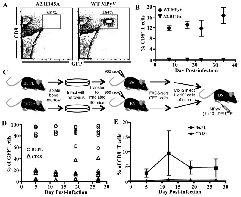
Q9:VP2.139-specific cells depend on intrinsic CD28 costimulation for expansion. 1 × 10⁶ Q9:VP2.139 TCR retrogenic GFP+ cells were transferred i.v. into B6 mice, which were subsequently infected with wild-type (WT) MPyV or A2.H145A. A. Representative dot plots of splenocytes in recipient mice at d 40 p.i., gated on viable lymphocytes. B. Frequency (± SD) of GFP+ Q9:VP2.139-specific CD8 T cells in blood over time. n = 3 mice. C. Experimental design: Q9:VP2.139 TCR retrogenic CD8 T cells derived from Thy1.1 B6.PL and B6.CD28−/− bone marrow were mixed 1:1, transferred to B6 mice, which were inoculated with WT MPyV. D. Frequency of Q9:VP2.139-specific CD8 T cells from B6.PL and CD28−/− bone marrow donors in blood over time. Dots represent individual mice. n = 3 mice. E. Frequency of Q9:VP2.139-specific cells from B6.PL bone marrow donors compared to cells from CD28−/− bone marrow donors in the blood over time following a co-transfer (± SD). n = 3 mice.

