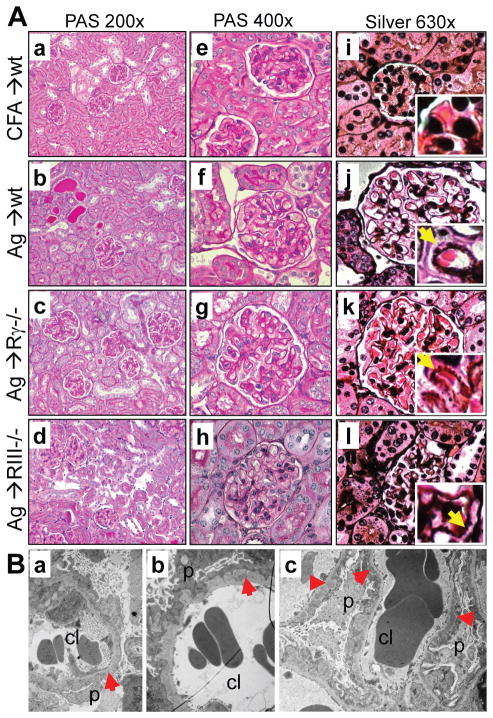
Figure 4. GBM thickening with formation of spikes and subepithelial electron-dense deposits are characteristic of membranous nephropathy.
A. Kidney sections from adjuvant-immunized control mice and wild type, FcγRIII−/− and FcRγ−/− mice immunized with rh-α3NC1, stained with PAS (a–h) or Jones silver (i–l) show GBM thickening but little glomerular inflammations or crescents, with occasional proteinaceous casts and mild interstitial fibrosis and inflammation. Jones silver stain shows GBM spikes (inset, arrows). Original magnification: 200× (a–d), 400× (e–h), 630× (i–l). B. Transmission EM shows subepithelial electron-dense deposits (red arrowheads), thickened GBM, and podocyte foot process effacement in α3NC1-immunized wild type (a), FcγRIII−/− (b) and FcRγ−/− (c) mice. Original magnification: 2850×.

