
Fig. 5.
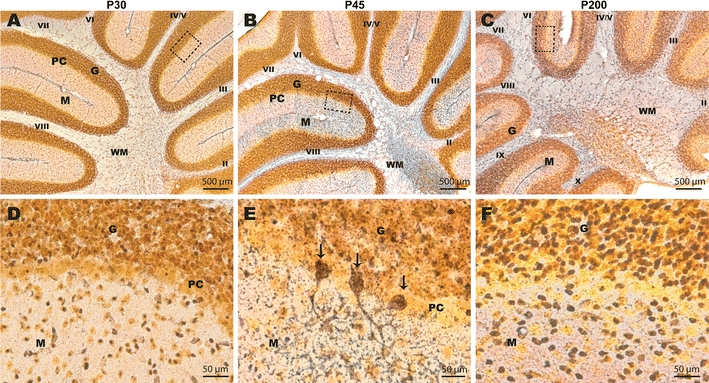
Silver impregnation of cerebellar sections in Cacna1a Purkinje cell-specific knockout mice. a, d No apparent neurodegeneration is seen in the cerebellum of PCα1KO mice at P30. b, e Silver-stained Purkinje cell bodies as well as dendrites and axons indicating dying cells in the cerebellum of PCα1KO mice at P45. Arrows indicate dying Purkinje cells in (e). c, f No Purkinje cells are present the respective areas. Silver-stained cells can be found in the granular and molecular layers indicating some granule and interneuron loss at P200–250. G granule cell layer, PC Purkinje cell layer, M molecular layer, WM white matter. Roman numbers indicate the cerebellar lobules. Dotted boxes in (a–c) correspond to regions shown in (d–f)