
Fig. 3.
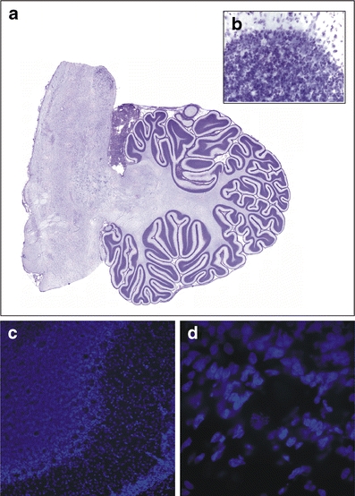
Photomicrographs of Nissl- and Hoechst-stained sections. a An example of a virtual slice of a Nissl-stained section of the cerebellum. b A 40× magnification example of a Nissl-stained section used to count granular cells. c–d An example of a Hoechst-stained section at a low (10×; c) magnification showing the different cerebellar layers and high (60×; d) magnification used to count Purkinje cells