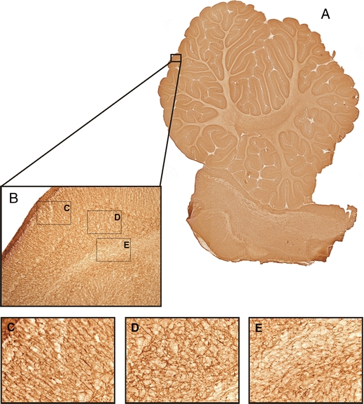
Fig. 4.

Photomicrographs GFAP. a An example of a virtual slice of GFAP-stained section of the cerebellum. b An example of a 10× magnification photograph of the GFAP staining showing the different layers of the cerebellum. c–e High magnification photographs (40×) of the molecular layer with the radial glial fibers (Bergmann glia) (c), the granular layer (d), and the white matter (e)
