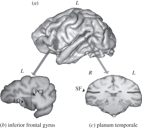
Figure 2.
(a) Three-dimensional reconstruction of a chimpanzee brain indicating the inferior frontal gyrus (IFG) and the planum temporale (PT). (b) 1 mm slice in the parasagittal plane with the IFG traced on the slice (FO, fronto-orbital sulcus; PCI, precentral–inferior sulcus). (c) 1 mm slice in the coronal plane with the sylvian fissure (SF) traced on the slice.

