
Fig. 4.
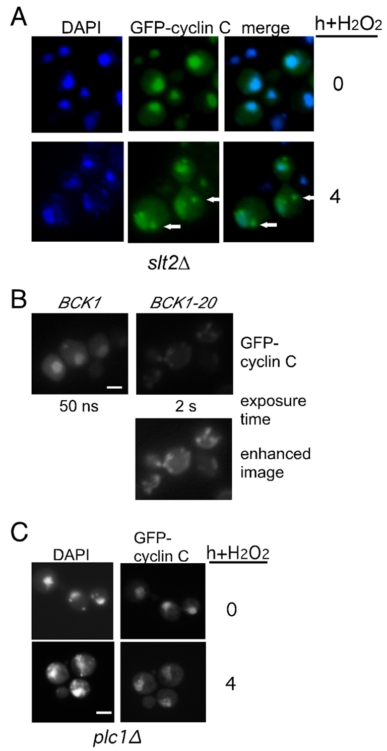
The Slt2p MAPK pathway is required for cyclin C cytoplasmic translocation. (A) An slt2Δ mutant strain (RSY1057) expressing GFP–cyclin-C (pBK1) was examined by fluorescence microscopy following heat shock (top panels) or 0.4 mM H2O2 treatment (bottom panels). Arrows indicate observed cytoplasmic foci. Nuclear location was followed with DAPI staining. (B) A wild-type strain (RSY10) expressing GFP–cyclin-C (pBK1) and either the constitutively active (BCK1-20) or the wild-type (BCK1) MEK kinase allele, were grown to mid-log-phase under non-stress conditions. The nuclei and GFP–cyclin-C were visualized as described for A. The exposure times for the top panels are indicated. The bottom panel was enhanced to more fully reveal the GFP–cyclin-C signal in the BCK1-20 expressing cells. (C) GFP–cyclin-C localization was monitored in a plc1Δ strain (RSY531) before and 4 hours following H2O2 exposure (0.4 mM). Exposure times were identical for visualizing the GFP–cyclin-C signal before and after stress application.