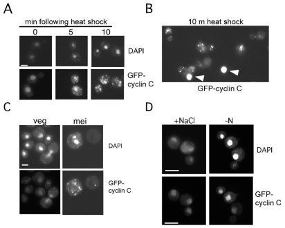
Fig. 5.
GFP–cyclin-C relocalizes in response to stressors that induce destruction. (A) A wild-type strain (RSY10) harboring pBK1 (GFP–cyclin-C) was subjected to heat shock (37°C) for the times indicated. Nuclear (DAPI) and GFP–cyclin-C signals were determined by fluorescence microscopy. (B) Comparison of GFP–cyclin-C nuclear and cytoplasmic foci signals following heat shock. Arrowheads indicate nuclear signals with the same exposure required to detect cytoplasmic foci. (C) Wild-type diploid culture (RSY335) harboring pBK1 was grown in synthetic acetate medium (veg) then transferred to sporulation medium for 6 hours (mei). The position of the nucleus (DAPI) and GFP–cyclin-C are indicated. (D) GFP–cyclin-C was visualized in a wild-type strain (RSY10) subjected to either high salt (0.4 M NaCl) for 30 minutes or starved for nitrogen (−N) for 2 hours. The position of the nucleus (DAPI) and GFP–cyclin-C are indicated. Scale bars: 5 μm.

