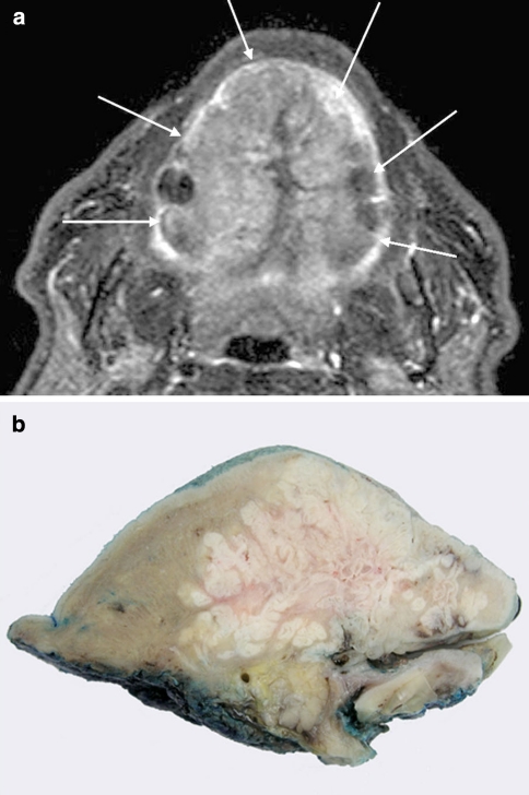
Fig. 1.
Radiological and macroscopic appearances. a T1 STIR axial MRI through oral cavity and tongue demonstrating extensive tumour (white arrows) involving the whole dorsal surface of the tongue. b Macroscopic appearance of a sagittal slice of the resection specimen demonstrating a complex endophytic pattern of infiltration

