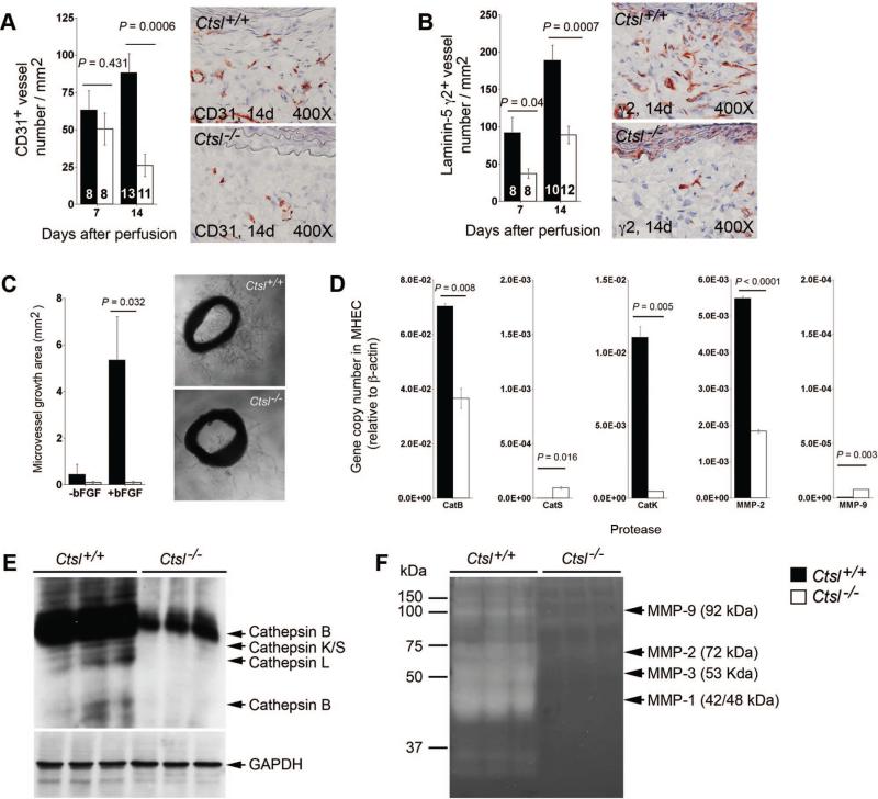
Figure 3.
Cathepsin L function in neovascularization in abdominal aortic aneurysms (AAA) lesions. CD31+ (A) and proangiogenic laninin-5 fragment γ2+ (B) microvessel numbers were reduced in AAA lesions from Ctsl–/– mice. The number of mice per group is indicated in each bar. Both measurements are from the entire lesion including adventitia and media. Aortic ring assay in vitro demonstrated impaired microvessel sprouting from Ctsl–/– mouse aortic rings with or without angiogenic factor bFGF (C). Representative images for panels A–C are shown to the right. RT-PCR showed reduced transcripts of cathepsins B and K, and matrix metalloproteinases (MMP)-2 in microvessel endothelial cells (MHEC) from Ctsl–/– mice compared with those from Ctsl+/+ mice (D). Cysteine protease active site labeling with JPM (E) and gelatin gel zymogram (F) demonstrated impaired cathepsin and MMP activities in MHEC from Ctsl–/– mice, respectively. GAPDH immunoblot was used for protein loading control. All data are mean±SE. P<0.05 was considered statistically significant; Mann-Whitney U test.

