
Fig. 11.
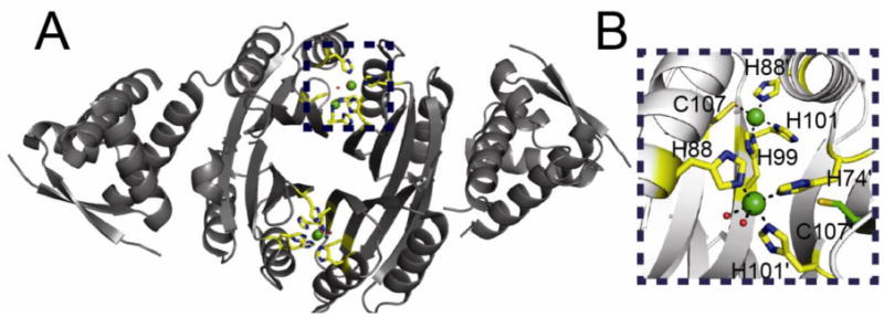
(A) Ribbon representation of the two pairs of Ni(II) binding sites of HpNikR solved at pH 5.6 [107]. Ni(II) ions are represented as green spheres. Metal binding residues are shown in stick representation with carbon atoms shaded yellow. (B) Close up of the two binding sites along the “top” of the NikR tetramer. Metal binding residues are shown as sticks with carbon atoms colored yellow. This view illustrates the two different coordination geometries that are found in this structure. The non-liganding Cys107 which forms part of the canonical square planar site (top site) is also shown in stick representation with carbon atoms colored green.