
Fig. 6.
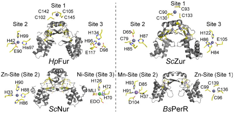
Illustration of multiple binding sites in the Fur family of repressor proteins. HpFur [64], ScZur [66], ScNur [75] and BsPerR [77] illustrate both the total number of different sites as well as the different coordination complexes that have evolved in a single protein family. Metal site designations are internally consistent and labeled sites 1, 2 and 3 according to the convention established for HpFur and MtZur [65] to facilitate comparisons. Sites 1, 2 and 3 in ScZur correspond to the sites previously identified at C, M and D, respectively.