
Fig. 7.
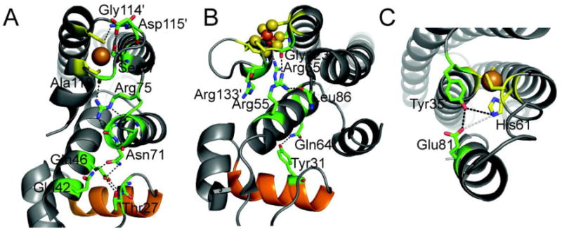
Putative hydrogen bonding network in the allosterically activated states of EcCueR [18] (A), EcSoxR (B) [90], and MtCsoR (C) [91]. Metal binding residues are shown in stick representation with carbon atoms shaded yellow. Key residues in the allosteric hydrogen bonding pathway are also shown as sticks with carbon atoms shaded green. The DNA recognition helix of the HTH DNA binding domain of CueR and SoxR is shaded orange. A native chemical ligation experiment carried out with MtCsoR is consistent with the coupling model shown [41]; the others have not yet been tested.