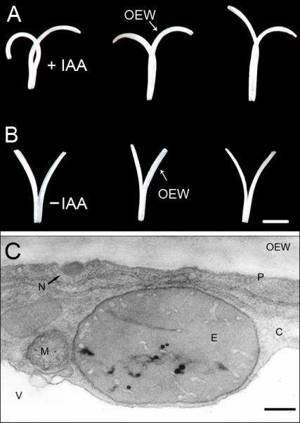
Fig. 3.

Effect of auxin (IAA, concentration: 10 μM) on the growth of split coleoptile sections, 15 mm in length, that were cut from 3-d-old etiolated rye seedlings (see Fig. 1). (A, B). After 24 h of incubation in the presence (+IAA) or absence (−IAA) of auxin, large differences are apparent in the three representative organ segments depicted here. IAA causes an inward curvature of the split halves via a selective promotion of the elongation of the growth-limiting outer epidermal wall. Transmission electron micrograph of a cross-section of an epidermal cell from an IAA-treated rye coleoptile segment (incubation time: 1 h; auxin concentration: 10 μM). (C). Note the electron-dense osmiophilic nano-particle in the periplasmic space between the plasma membrane and the cytoplasm. C = cytoplasm, E = etioplast, M = mitochondrion, N = osmiophilic nano-particle, OEW = outer epidermal wall, P = plasma membrane, V = vacuole. Bars = 1 cm (A, B); 0.5 μm (C).
