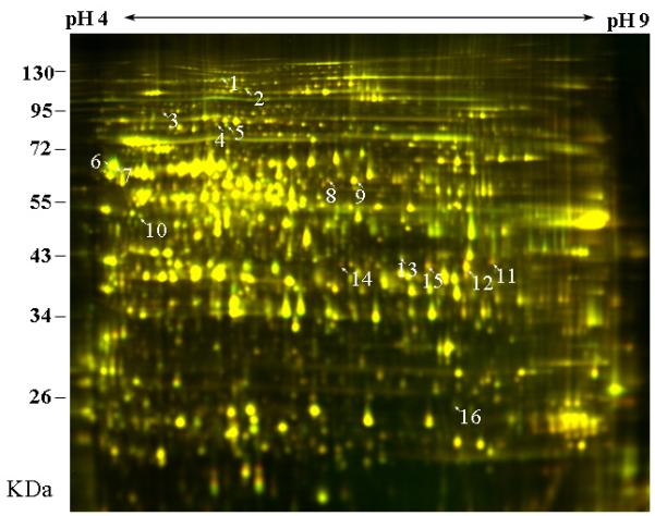
Fig. 4.

Effects of auxin (IAA) on the proteome of rye coleoptile, as revealed with two-dimensional DIGE analysis of microsomal proteins. Segments, 15 mm in length, were incubated for 2 h in the presence or absence of auxin (± IAA, concentration: 10 μM). Thereafter, membrane-associated proteins were extracted and labelled with Cy3 dye (−IAA) or C5 dye (+IAA). The labelled microsomal proteins were mixed and separated on the first dimension with 24-cm, pH 3 to pH 10, NL IPG strips, and thereafter on a second dimension with a 10% polyacrylamide SDS-PAGE gel. The images were analysed with a Typhoon Trio scanner and superimposed. Protein spots up-regulated in the presence of IAA appear in red, and those down-regulated in green. Spots that are unaffected by the hormone are yellow.
