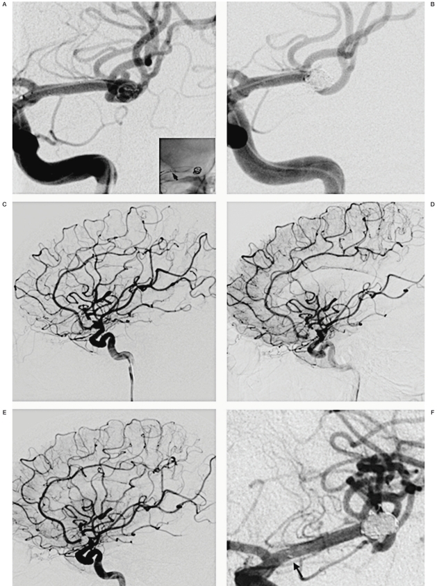
Figure 2.
A) DSA magnified view demonstrates a wide-necked bilobular left MCA bifurcation aneurysm. A native view in the right lower corner shows the proximal end of the stent (a smaller arrow). B) DSA magnified view demonstrates Raymond Class I occlusion of the aneurysm. C) DSA lateral view demonstrates normal fillings of left MCA immediately after the procedure. D) DSA lateral view demonstrates delayed fillings at left central cortical branch and posterior parietal branch arteries at 18 hrs after the treatment. E) DSA lateral view demonstrates complete recanalization and normalized transit time of those branch arteries next day. F) DSA magnified view demonstrates persistent occlusion of the aneurysm and the widely patent stent at 6 months. Arrow indicates the proximal end of the stent.

