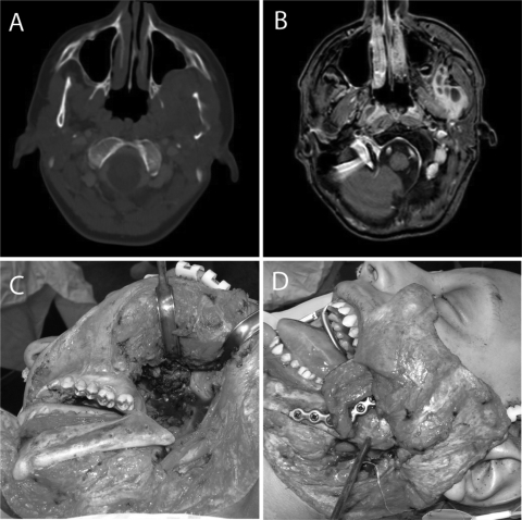
Figure 1.
A 14-year-old boy with Ewing's sarcoma. Axial computed tomography (A) and magnetic resonance (B) images show the tumor involving the left mandible and infratemporal fossa. Tumor resection included a segmental mandibulectomy (C), and reconstruction was preformed with a fibula free flap and titanium plate (D).

