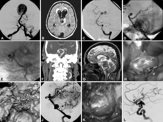
Figure 1.
Intraoperative and angiographic images of cerebral aneurysms with complex features, including examples of giant size (A), intraluminal thrombosis (B), complex configuration (C), difficult access location (D), previous treatments (E), calcification of the aneurysm wall (F), embedding on surrounding tissues (G), blister-like aneurysm (H), aneurysm involving parent artery (I), branch arising from aneurysm (J), broad neck (K), and fusiform aneurysm (L). (Reprinted with permission of Mayfield Clinic.)

