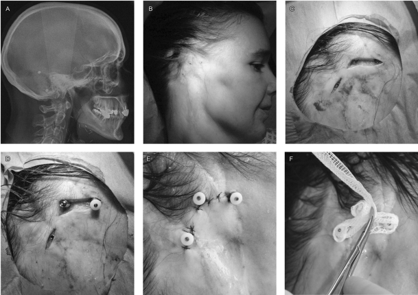
Figure 2.
Skin incision and fixture positioning. A 39-year-old woman who underwent resection of a cystic adenoid carcinoma of the parotid followed by irradiation of 50 Gy. (A) Lateral cephalogram showing four fixtures, three upon the mastoids for prosthesis, one posterior for bone anchored hearing aid. (B) Preoperative localization of implants. (C) Skin incisions showing implants with their cover screw. (D) Placement of transcutaneous fixtures, the posterior implant has the cover screw removed, the middle one has the transcutaneous fixture and the anterior one has the protective cap on top of the fixture. (E) Sutured skin closure to obtain good contact between fixtures and the skin, limiting subcutaneous tissue exposure. (F) Dressing with iodoform gauze, to prevent granulation tissue (even on irradiated tissues)

