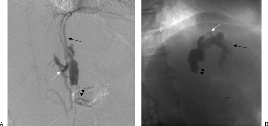
Figure 4.
Type B venous outflow. (A) With the occlusion balloon (arrowheads) positioned at the base of the gastrorenal shunt, balloon-occluded retrograde venography shows filling of multiple “leaking” collateral veins including inferior phrenic (black arrow) and perivertebral veins (white arrow).(B) After the occlusion balloon (arrowheads) was advanced beyond the “leaking” collateral veins, venography through a microcatheter (white arrow) now shows opacification of the gastric varices (black arrow).

