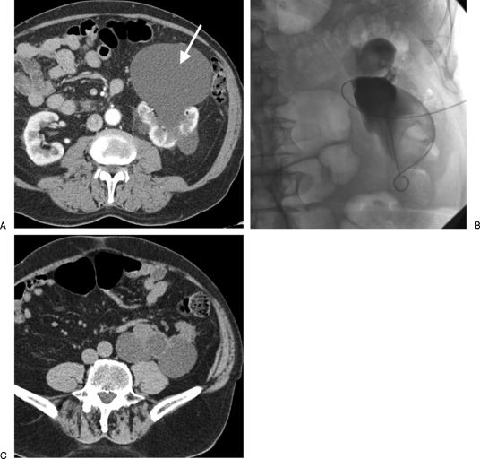
Figure 1.
(A) A 78-year-old man with pain and pressure caused by a large anterior left renal cyst (arrow). (B) A catheter was inserted and the cyst drained. This image shows the infolding of the cyst as a large volume of fluid was drained. The cyst was then sclerosed with alcohol that was later aspirated in a single session. Note the catheter was inserted from an anterior approach given the large size of the cyst, thus the orientation of this image. (C) Follow-up computed tomography 9 months later shows no recurrence of the anterior cyst that was sclerosed although the posterior cysts have grown in size.

