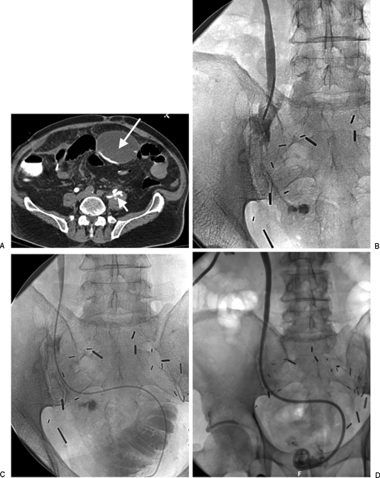
Figure 4.
(A) The patient is postcystoprostatectomy with ileal neobladder formation with leak from a ureteral injury. The computed tomography scan shows both local extravasation (short arrow) from the ureter as well as tracking of contrast into a more remote anterior urinoma (long arrow). (B) Direct injection in the ureter from a catheter introduced through a percutaneous nephrostomy show the periureteric extravasation. (C) An angiographic catheter was able to be manipulated past the ureteral leak into the neobladder. An internal-external nephroureteral catheter was then placed to provide diversionary drainage. (D) Follow-up nephrostogram just 2 weeks later shows no further extravasation from the ureter.

