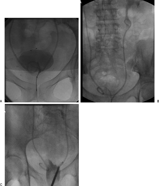
Figure 6.
Multiple images from different patients showing the steps of a double-J stent change. (A) An MPA-guiding catheter used to distend the bladder with dilute contrast and guide the snare to grab the distal end of the stent. (B) The distal end was snared and pulled out through the urethra. Notice the cephalad end of the double-J stent is maintained high within the ureter to avoid losing access to the ureter. The stent was injected with contrast to visualize the pelvicaliceal system to help in the proper positioning of the new stent. (C) Another patient where a McGill forceps was used to grab the stent.

