
Figure 7.
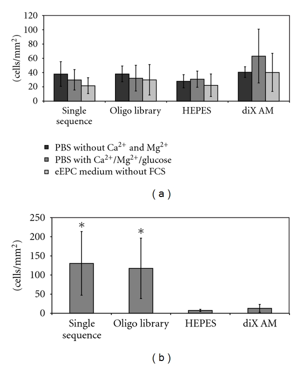
Influence of FCS on cell adhesion on oligonucleotide coated diX AM surfaces. (a) Samples were incubated with 0,05 ∗ 106 eEPC cells for 2,5 h under dynamic conditions in medium or buffer without FCS. Cell number was counted in microscopy images (n = 6). ANOVA tests revealed no significant difference between the groups. (b) Samples were preincubated with eEPC Medium containing FCS for 2 h. Afterwards 0,05 ∗ 106 eEPC cells were applied for 2,5 h under dynamic conditions in medium without FCS. Cell number was counted in microscopy images (n = 6). Groups marked with ∗ are significantly different to HEPES and diX AM samples (P < 0.05).