
Figure 9.
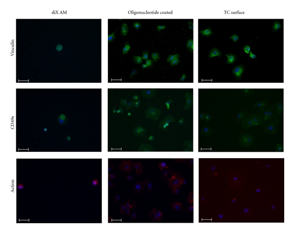
Staining for Vinculin, CD49e and F-Actin of HUVECs after 1 h hour incubation in FCS containing medium under dynamic conditions. Samples were incubated with 0,05 ∗ 106 cells in medium containing FCS. Nonadherent cells were removed before staining. Scale bar equals 50 μm. Nuclei are stained with DAPI (blue).