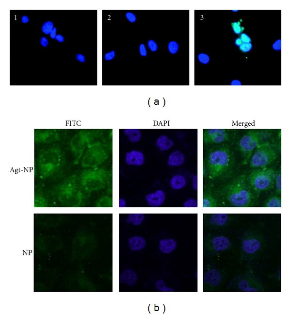
Figure 9.

Targeted nanoparticles are promising for future in vivo gene delivery approaches. (a) PSMA-targeted PLGA-based microparticles enter LNCaP (PSMA+) PCa cells. Untreated control (1), after 30 min of exposure to nontargeted FITC-loaded (2), and targeted FITC-loaded (3) MBs. Cell nuclei were stained with Hoechst (blue). The number of green-positive cells per field was significantly different from that of nontargeted MBs. Reprinted from [64] with permission from American Chemical Society. (b) Confocal fluorescent scanning microscopy images detecting cellular uptake of MUC-1 targeted Aptamer conjugated NPs (top row) or NPs (bottom row) in MCF-7 cells. Green fluorescent FITC was encapsulated in Apt-NPs and NPs. The nuclei were stained blue with DAPI. The right column showed the merged images of the FITC and the DAPI channels. MCF-7 cells were exposed to FITC-encapsulated Apt-NPs or NPs at 100 μg/mL for 2 hours. Reprinted from [66] under the terms of the Creative Commons Attribution License.
