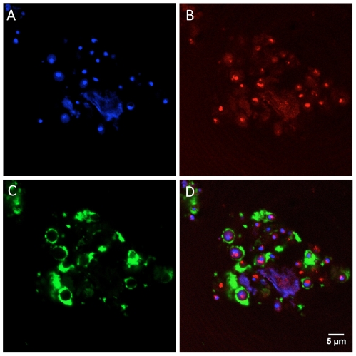
Figure 6. Binding of biotinylated N10K to Candida albicans cells.
A. Cells stained with DAPI; B. Phalloidin-rodamine stained cells. C. Biotinylated N10K-treated cells stained with streptavidin-fluorescein isothiocyanate. Fluorescence is seen at the cells' periphery, mainly at the cell wall. D. Merge produced by the superposition of the red and green fluorescence outputs of the same cells. The peptide ligand does not colocalize with F-actin.

