Figure 1.
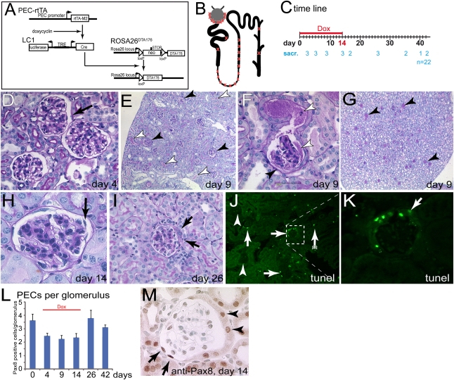
Transgenic model for inducible ablation of PECs. (A) Schematic of the transgenes. The reverse tetracycline-inducible transactivator (PEC-rtTA) is expressed in PECs. Upon administration of doxycycline, Cre recombinase is transiently expressed from the second transgene (LC1). This activates expression of an attenuated DTA (DTA176). (B) Schematic of expected cellular ablation within the nephron. The transgenic PEC-rtTA is active in PECs, the loop of Henle, and the thick ascending limp (as indicated with an X). In addition, single scattered cells are targeted within the proximal tubule and the collecting duct. (C) Study timeline. Animals were treated with doxycycline for 14 days and killed at the indicated time points (numerals indicate the number of sacrificed animals per time point). (D through I) PAS-stained paraffin section of the kidney at different time points. (D) On day 4, no histomorphological abnormalities were observed in PECs (arrow) or elsewhere within the kidney (female). (E) On day 9, protein was observed within the Bowman’s space of a fraction of the glomeruli (black arrowheads). Protein-filled tubuli are marked by white arrowheads. (F) Protein has leaked into the primary urine (white arrowheads). Parts of the inner aspect of Bowman’s capsule are covered by fuzzy material (black arrowhead). (G) Within the renal papilla, a fraction of the tubules contain protein (arrowheads; mice in E through G are male). (H) On day 14, PECs appear partly detached (arrow, female). (I) Late time points are characterized by extracapillary proliferations (i.e., cellular crescents, arrows, 10-week-old female mice). (J) TUNEL assay 9 days after induction. A fraction of the glomeruli contain TUNEL-positive PECs (arrows), whereas other glomeruli do not show any TUNEL-positive cells (arrowheads). Scattered tubular TUNEL-positive cells could be observed (arrow with tails). (K) Higher magnification of inset in J. Cells lining the inner aspect of Bowman’s capsule contained DNA fragments. Negative control not shown. (L) Pax8-positive cells quantified per glomerulus at different time points (n=3 per time point, 50 glomeruli per mouse). Overall, approximately 30% of all PECs were ablated. (M) Representative image of a glomerulus. Pax8-positive oval nuclei of cells with a flat cytoplasm were counted as PECs. Tubular cells lining Bowman’s capsule were not counted as PECs (arrowheads, male). Dox, doxycycline; sacr, sacrificed.

