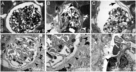
Figure 2.
Ultrastructural analysis of subtotal PEC ablation. (A) Normal glomerulus 9 days after induction. (B) Cellular debris (black arrowhead) and protein within Bowman’s space on day 9. Remnants of PECs line Bowman’s capsule (white arrowheads), also within an adjacent glomerulus (arrow). (C) PECs have partially detached from Bowman’s capsule (arrow) (A through C, semi-thin sections). (D and D′) TEM images of a glomerulus 9 days after induction of PEC ablation. A viable PEC is marked by a white arrow. The remaining PECs are disintegrating (black arrowheads; D′ shows a higher magnification of the inset in D). (E) In this higher magnification, the apical plasma membrane of the PEC has disappeared. The cellular contents including the organelles are in direct contact with Bowman’s space (arrowhead). Bowman’s capsule (i.e., the PEC basement membrane) is wrinkled but still intact (white arrow, all animals aged 8 weeks; female mice are shown in A, C, and D, and male mice are shown in B and E).

