Figure 2.
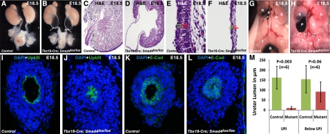
Smad4 inactivation in the ureteral mesenchyme leads to hydronephrosis and UPJ obstruction. (A and B) Smad4 inactivation in ureteral mesenchyme results in severe bilateral hydronephrotic kidneys having kinked ureter at UPJ in mutants compared with normal ureter phenotype in controls. (C and D) Severe renal pelvis retention and fluid pressure resulted in decreased thickness of epithelium and smooth muscle layer at the UPJ of mutants compared with wild type. A higher magnification of the sagittal section of the pelvis shows a thinner layer of epithelium and mesenchyme in (F) mutant compared with (E) control. Ink is injected in the pelvis of right kidney of control and mutant mice. (G) Ink injected in the pelvis of control kidney reaches to the bladder (evidenced by accumulation of ink in the bladder), whereas (H) ink cannot pass through the UPJ in mutants and reach to the bladder (shown by the absence of ink in the ureter and bladder). White arrows indicate the UPJ. Immunostaining of control and mutant sections at UPJ by anti-UpkIII and anti–E-Cad antibodies shows narrow ureteral lumen in (J and L) mutants compared with (I and K) normal lumen in controls. (M) Statistical measurement (two-tailed t test) of lumen diameter in the mutants versus controls shows more severe constriction of ureter lumen at UPJ (P=0.003, n=6) than below UPJ (P=0.06, n=6). B, bladder; Epi, epithelium; K, kidney; Mes, mesenchyme; P, pelvis; T, testis; U, ureter.

