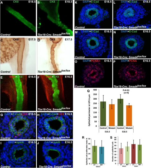
Figure 4.
Loss of Smad signaling specifically in the ureteral mesenchyme also affects ureteral epithelium. (A–F) Staining of control and mutant ureteral epithelium by anti-CK8 antibody shows (B, D, and F) irregular epithelium throughout the length of the mutant ureter compared with (A, C, and E) normal smooth outlined epithelium in control ureters. Immunostaining of ureter epithelium by CK5 and UpkIII to label the epithelial cells in the basal layer and on the luminal surface, respectively. In addition, E-Cad stains all the epithelial cells. (G–J) No significant difference in the cellular composition between mutants and controls was found. Immunostaining by an anti–E-Cad antibody targeting the epithelium shows narrow ureter lumen at the (L) mutant UPJ compared with (K) normal ureteral lumen of control at E18.5, which is not observed at (M and N) E16.5. (Q) Epithelial perimeter (in micrometers) at E16.5 was controls, 270.35±69.06 (n=4); mutants, 241.55±50.78 (n=4); P=0.45. Epithelial perimeter (in micrometers) at E18.5 was controls, 301.31±46.28 (n=4); mutants: 229.7±20.08 (n=4); P=0.01. Immunostaining of (O) control and (P) mutant cross-sections by antiproliferation cell nuclear antigen (PCNA) antibody indicates no difference in the cellular proliferation. (R) Metric data analysis (two-tailed t test) also indicates no difference in the proliferation index of epithelial cells of mutant (34±13, n=4) and control (36±11, n=4) ureters at E16.5 (two-tailed t test, P=0.83) and (S) consequently, no difference in the epithelial cell number at E16.5 (controls, 51.33±17.18 [n=4]; mutants, 57.38±15.13 [n=4]; P=0.06) and E18.5 (controls, 65.22±13.9 [n=4]; mutants, 63.33±17.22 [n=4]; P=0.50). CT, control; MT, mutant.

