Figure 3.
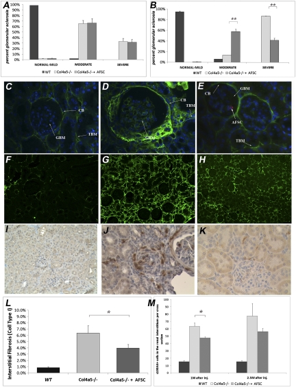
AFSC injections significantly ameliorate glomerular and interstitial fibrosis in AS mice. AFSC injection ameliorated glomerular sclerosis (A and B). Morphometric analysis of percentage of sclerotic glomeruli in Col4a5−/− mice injected with AFSCs (n=10) at 1.5 months after birth and killed at 1 (A) and 2.5 months (B) after injection and similarly for noninjected Col4a5−/− mice (n=10) is shown. Using periodic acid-Schiff staining, 50 randomly selected glomeruli per mouse were scored according to the severity of fibrosis as follows: normal-mild (0%–33%), moderate (33%–66%), and severe (66%–100%). As shown by graphs, the injected mice present less sclerotic glomeruli than their noninjected siblings at 2.5 months after injection. AFSCs reduced COL4α1 deposition within the glomeruli (C–E). Injected mice showed less glomerular deposition of COL4α1 compared with noninjected mice. Representative pictures show COL4α1 distribution (green) in wild-type mouse (C, ×40), noninjected Col4a5−/− mouse killed at 4 months after birth (D, ×40), and Col4a5−/− mouse injected with AFSC at 1.5 months after birth and killed at 2.5 months after injection (E, ×40). Rare AFSC labeled red with CM-DiI were seen inside the glomeruli of injected mice (arrow, E) (BC, Bowman capsule; TBM, basal membrane of the tubules; GBM, glomerular basement membrane). Injection of AFSC diminished the deposition of extracellular matrix in the interstitium, as shown by quantification of collagen I (F–H and L). In the representative picture of injected mice (H, ×20; n=10) the presence of collagen I is clearly less than in the noninjected mice (G, ×20; n=10) and is much more similar to that of the wild type (F, ×20; n=10). These data were confirmed by quantification of staining. Graph in L compares collagen I accumulation per cross-section in all experimental groups analyzed using HistoQuest software, revealing a statistically significant difference at 2.5 months after injection. Injection of AFSC also reduced the number of S100A4-positive cells (I–K and M). This marker is mostly expressed by cells undergoing myofibroblast transformation and by cells actively producing collagen during the progression of fibrosis, including endothelial cells and macrophages. In treated mice (I, ×20), the number of S100A4-positive cells in the interstitial space is reduced compared with their nontreated siblings (J, ×20). The graph in M represents the change in number of S100A4-positive cells per cross-section in mice injected (n=10) at 1.5 months after birth and killed at 2.5 and 4 months of age versus noninjected (n=10) mice of the same age. All values are presented as mean ± SEM (*P<0.05). WT, wild type.

