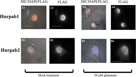
FIGURE 9.
Hnrpab isoforms appear in the cytoplasm of mature neurons. Primary hippocampal neurons from E18 Hnrpab+/− mouse brains were dissociated and plated on coverslips and then maintained in culture for 8 d before infection with recombinant LVPs to express Flag-epitope tagged either Hnrpab1 (A) or Hnrpab2 (B). On 15DIV cultures were treated with 50 μm of glutamate (panels labeled 3 and 4) or mock treated (panels labeled 1 and 2) for 10 min and then glutamate removed and incubation continued for 6 h. These were fixed and processed for immunostaining using anti-Flag epitope antibodies, and DAPI for the nucleus. Pseudo-colored Flag immunofluorescence (orange) merged with DAPI (blue) and DIC images (panels labeled 1 and 3) are shown next to the Flag immunofluorescence images alone (panels labeled 2 and 4). Scale bars represent 20 μm.

