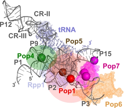
FIGURE 3.
UV-induced RNA–protein crosslinks mapped onto the outline of the RNA component of RNase P. The outline is based on the crystal structure of bacterial RNase P (Reiter et al. 2010). (Gray) RNase P RNA; (light gray) tRNA product; only the RNA elements that are universally conserved from bacteria to eukaryotes are shown. (Solid spheres) The locations of the identified crosslinking sites for individual proteins. (Red) The location of the Pop1 crosslink; (green) the location of the Pop4 crosslink; (brown) the location of the Pop5 crosslink (carried over from RNase MRP); (magenta) the locations of the Pop7 crosslinks. Corresponding approximate locations of proteins are shown as semitransparent in matching colors. The structures of Pop6 and Pop7 are modeled according to Perederina et al. (2010b); Pop5 and Rpp1 are modeled according to Perederina et al. (2011). RNA structural elements are marked according to the nomenclature used in Figure 2B.

