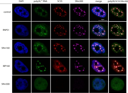
FIGURE 6.
Subcellular distribution of poly(A)+ RNAs in cells where the expression of nuclear speckle proteins was repressed by RNAi. FISH analyses of HeLa-TO cells transfected with the indicated siRNAs (poly(A)+ RNA, green) are displayed, together with immunocytochemistry of SRSF2 (SC35) (red), SRm300 (cyan), and DAPI staining of DNA (blue).

