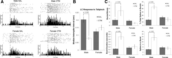
Figure 2.
Tail-pinch elicited firing of single LC neurons. A, B, The median interspike interval following tail-pinch was shorter (demonstrating faster firing) in male rats neonatally exposed to CTM (0.079 ± 0.035 s; n = 15) compared to male saline (SAL) (0.204 ± 0.135 s SD; n = 47), female SAL (0.124 ± 0.111 s SD; n = 38), and female CTM (0.147 ± 0.102 s SD; n = 12) animals, verified with a significant sex by drug exposure interaction (F(1,108) = 8.83, p = 0.004) and a simple main effect of drug exposure for males (F(1,108) = 14.052, p < 0.001). Perievent rasters and corresponding histograms are of representative animals with the vertical dashed line illustrating the onset of tail-pinch. The raster plots illustrate the trial-by-trial responses of a single LC neuron to tail-pinch. Histograms are in 10 ms bins converted to spikes/s to show the average response of the cell over ∼5 min. C, Burst analysis of single LC neurons demonstrated a significant increase in (1) the number of bursts per minute, (2) the percentage of total spikes categorized within a burst, (3) mean burst duration, and (4) the number of spikes within a burst of male rats exposed to CTM compared to all other groups. Error bars, SEM.

