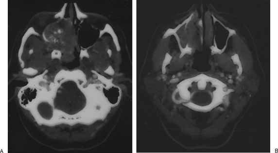
Figure 3.
Axial computed tomography scan with contrast demonstrates a large tumor of the right maxillary sinus with extension through the posterior wall to involve the pterygopalatine fossa and infratemporal fossa (A). There was no erosion of the anterior wall of the maxilla and no involvement of the premaxillary soft tissues (B).

