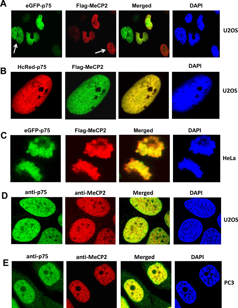
Figure 4. Nuclear co-localization of LEDGF/p75 and MeCP2 by confocal micrscopy.
U2OS cells were transiently co-transfected with plasmids encoding Flag-MeCP2 and eGFP-LEDGF/p75 (A), or the combination of HcRed-LEDGF/p75 and Flag-MeCP2 (B). HeLa cells were transiently co-transfected with plasmids encoding Flag-MeCP2 and eGFP-LEDGF/p75 (C). White arrows in panel A point to cells that were stained with eGFP-LEDGF/p75 but not with Flag-MeCP2, or viceversa, ruling out the possibility of bleeding from one channel into the other. Ectopically expressed MeCP2 was detected 48 h post-transfection using anti-Flag antibodies and visualized with FITC-labeled secondary antibody. Endogenous co-localization of LEDGF/p75 and MeCP2 was observed in U2OS and PC3 cells after co-incubation with human anti-LEDGF/p75 and rabbit anti-MeCP2 antibodies, followed by detection with corresponding secondary antibodies. Nuclei were stained with DAPI, and fluorescent signals were analyzed by confocal microscopy.

