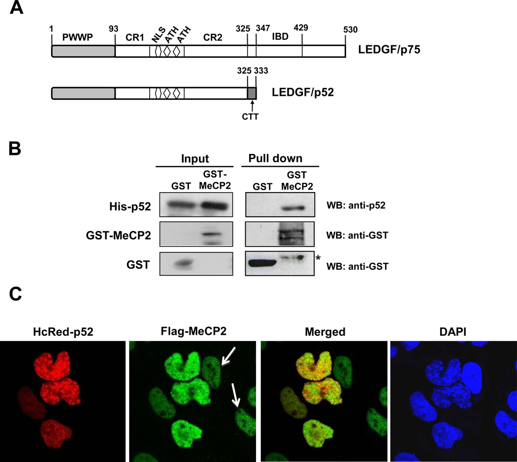
Figure 5. LEDGF/p52 interacts with MeCP2.
(A) Schematic domain structure of LEDGF/p75 and p52. (B) Pull down assay was performed as described in the legend of Fig. 2A using recombinant His-LEDGF/p52. *Denotes degraded MeCP2. Protein input was determined by immunoblotting of whole cell extracts. (C) LEDGF/p52 partially co-localizes with MeCP2 in the cell nucleus. White arrows point to cells that were stained with Flag-MeCP2 but not with HcRed-p52, ruling out the possibility of bleeding from one channel into the other. Co-localization assay was performed as described in the legend of Fig. 4.

