
Fig. 7.
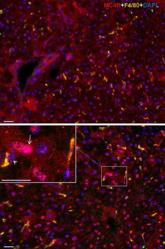
Double immunofluorescence staining with polyclonal antibody directed against MC4R (red) and F4/80 (green) followed by fluorescent immunodetection in sections of mouse liver during APR. Upper control liver, lower 6 h after TO treatment. Inset shows higher magnification. White arrow indicates the hepatocytes positive for MC4R and white arrowhead indicates the Kupffer cells positive for MC4R and F4/80. Results show the representative picture of three animals and six slides (original magnification, ×200, scale bar 20 μm)